
Radiologists and parents have something major in common — we are always taking pictures of our children.
As a parent, your camera roll is probably filled with the birthday parties, milestones, and fun moments you’ve captured. Our pictures aren’t quite as pretty, but they are just as important. The pictures we take show us the inside of your child’s body so that we can look for signs of disease or injury and help your child heal.
Make An Appointment
Your child will need an order from a provider to schedule a radiology procedure. Once the order is placed, call 402-955-6799 to schedule the procedure. Take advantage of our offsite clinics to save yourself a trip to the hospital. Please call ahead, as not all studies can be completed offsite.
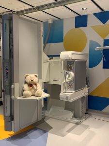
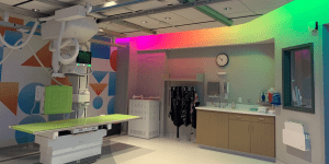
Radiology involves a lot of equipment, odd noises, and sitting still — three things that kids don’t always enjoy. But here at Children’s Nebraska our pediatric-trained specialists know exactly how to coax kids through a radiology appointment. We understand the joys and quirks of working with children. We know when a child needs the distraction of TV or music, or simply needs a hand to hold.
We are also very aware of what children don’t need, which is more radiation than is absolutely necessary to get the most accurate images possible. We use “child size” doses of radiation to decrease long-term cancer risks, and provide alternative tests that do not involve any radiation.

- Using small, child-size equipment reduces the risk of your child being overexposed to radiation.
- No radiation remains in your child’s body after X-rays or CT scans.
- The amount a child is exposed to during a test is calculated for each child on an individual basis, based on their size and weight.
What Sets Children’s Apart?
Radiology is an important step in your child’s treatment plan. An accurate reading (interpretation) of your child’s pictures means a correct diagnosis. In the same way, an incorrect reading of pictures can lead to an incorrect diagnosis. This can lead to the wrong course of treatment, and being unable to solve the problem at hand.
We won’t let that happen.
Our Radiology staff has extensive experience working with one patient population: children. We excel at making them feel comfortable in situations that can be a little unsettling, and we collaborate with referring physicians to determine the most effective way to take pictures of your child for an accurate diagnosis.
Our radiologists are fellowship-trained pediatric radiologists. They focus only on pediatric images and are specifically trained in diseases that are seen in children. All our imaging protocols are built specifically for pediatrics. Our radiologists have over 65+ years of combined experience reading pediatric studies and have been recognized nationally for their efforts to minimize radiation exposure while providing diagnostic results.
Additionally, Children’s Radiology team has received national recognition for:
- Following the recommendations of Image Gently®, a program of the Alliance for Radiation Safety in Pediatric Imaging. The campaign goal is to promote radiation protection in the imaging of children. Children’s has been embracing their guidelines, and their motto of keeping radiation doses “as low as reasonably achievable,” for the past decade.
- Stepping forward as a national advocate for the use of sonography to diagnose pediatric appendicitis. This is another element of our continuing effort and dedication to minimizing a child’s radiation exposure.
- Offering a low dose, 3D X-Ray imaging system — the EOS system® — that uses 50 to 80% less radiation than a typical X-Ray. This enables us to provide As Low As Reasonably Achievable (ALARA) imaging, which means it limits your child’s exposure to radiation to the lowest and safest amount possible to still get the results we need. With EOS, our physicians can make an informed diagnosis and develop treatment plans for your child’s spine, hip, or knee condition. Children’s is the only hospital in Nebraska offering EOS imaging.
- Being honored with a Health Devices Achievement Award by the ECRI Institute, a non-profit organization which researches the best approaches to improving patient care. Children’s was recognized nationally for its innovation in developing an easy and logical approach to ensure the proper use of diagnostic and radiological imaging, and allowing medical staff to be more efficient and accurate in their testing choice.
- Our CT and MRI departments being recognized by the American College of Radiology (ACR) as an accredited facility for offering gold standard of care in pediatric medical imaging.
“The technologists were great at talking with my child and explaining the process of his tests so my child knew what to expect and was comfortable at all times. They made sure I was aware of the process and how it would go, and kept me up to date on the progress of the test.”
—Parent of Children’s Nebraska patient
Services We Provide
-
3D Visualization Lab
Our 3D Visualization Lab uses the latest in cutting-edge technology to provide the best care for our patients. By combining advanced imaging practices with 3D modeling, our physicians are better prepared for the most difficult cases by creating a virtual model that can be printed in 3D.These 3D models allow physicians, patients, and patients’ families to fully understand what will be done during a procedure, as well as increased staff knowledge of each case. This pre-surgical planning method leads to better outcomes for our patients.
-
Bone Densitometry (DEXA)
Bone densitometry (DEXA) is used to test bone mineral density — a measurement of the amount of calcium and other minerals in the bone. This measurement can help us find decreased bone mass, a condition in which the bones are brittle and can break easily.
Usually, we use DEXA to diagnose osteoporosis (a condition that causes the bones to become thin and fragile), and determine a child’s risk of breaking bones. DEXA examines bones of the spine, pelvis, lower arm, and thigh.
During the procedure, the DEXA machine sends X-rays through the bones being examined. Some of the energy from the x-ray is absorbed by the bone, and the rest of the energy is absorbed by soft tissue. Special DEXA software calculates the amount of energy absorbed by the bone and tissue, and computes bone density measurements.
Your child will be exposed to a very small amount of radiation. Since your child’s safety is our priority, we follow recommendations from the Image Gently® Campaign — a program from the Alliance for Radiation Safety in Pediatric Imaging that promotes X-ray safety measures specifically geared towards protecting children from radiation.
-
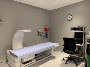
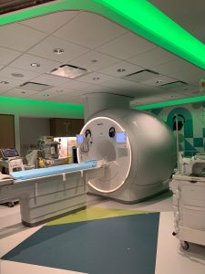
CT (Computer tomography) Scan
During a computed tomography scan (also called CT or CAT scan), your child will lie on a narrow table that slides into a part of the scanner that is shaped like a doughnut. An X-ray beam moves in a circle around the body and takes detailed pictures of an organ or structure in the body. CT pictures are much more detailed than traditional X-ray pictures, especially in blood vessels and soft tissues.
For certain CT scans, your child may need to receive contrast — a substance that’s taken either by mouth or injected directly into the body via an IV, and allows us to see the organ or tissue being studied more clearly. There are times your child will receive contrast both orally and through an IV.
Unless your child is given a sedative or anesthesia, you will be allowed to stay with them while they’re getting the scan.
CT scans do involve the use of radiation, but our CT doses are “child size.” That means the doses are carefully calculated to decrease long-term risks of radiation. We also only use CT scans whenever absolutely necessary. Whenever possible, we use ultrasound or magnetic resonance imaging (MRI) tests to minimize radiation exposure.
When we do need to use CT scans, we follow recommendations from the Image Gently® Campaign — a program from the Alliance for Radiation Safety in Pediatric Imaging that promotes X-ray safety measures specifically geared towards protecting children from radiation.
-
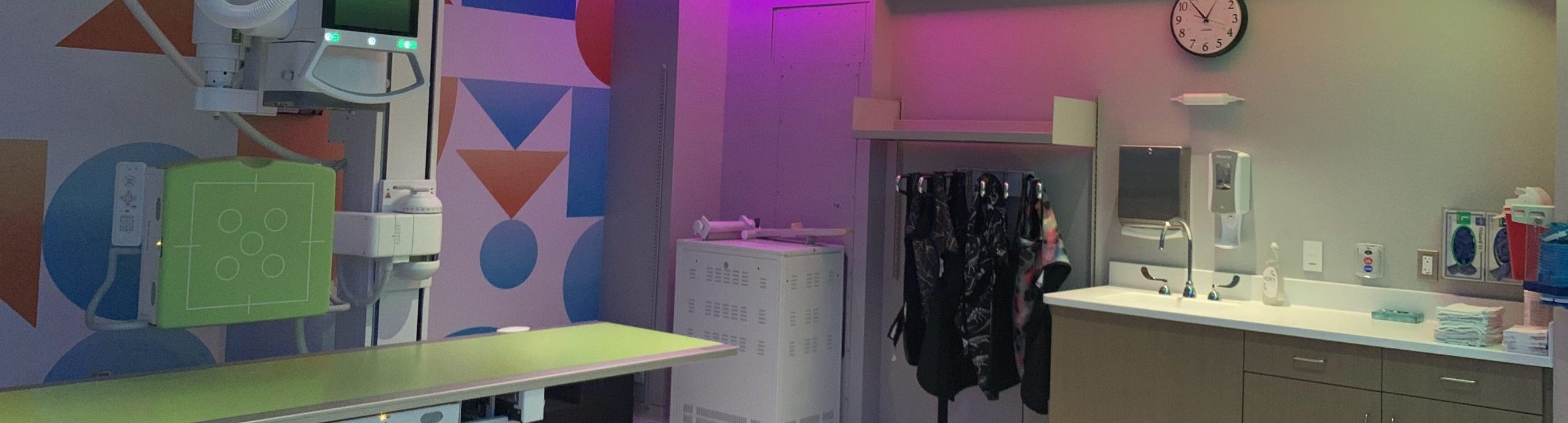
Digital Fluoroscopy
Digital fluoroscopy uses X-rays to take detailed, moving images of organs and body structures, such as the intestines, bladder, heart muscle, and stomach. Fluoroscopy produces a “live” look at what’s going on inside the body- like a video camera using X-rays.
In some cases, your child may need to receive contrast — a substance that’s taken either by mouth or injected directly into the body — that allows us to see the organ or tissue being studied more clearly.
There are many types of tests that can be performed using digital fluoroscopy. For example, we may use it for a barium enema, which is a test that looks for disease in the large intestine (colon). Or, we may use fluoroscopy to perform a voiding cystourethrogram, or VCUG — a test that evaluates the kidneys, bladder, and ureters (tubes connecting the kidney and bladder), and can diagnose reflux of urine from the bladder to the kidneys.
Fluoroscopy exams can be difficult to complete, even for adults. That’s why we’ve taken steps to ease the process for our young patients. We utilize tools for distraction (like electronic tablets, toys, and music), and are always there to hold your child’s hand.
Since fluoroscopies use X-rays, your child will be exposed to radiation. However, we follow recommendations from the Image Gently® Campaign — a program from the Alliance for Radiation Safety in Pediatric Imaging that promotes X-ray safety measures specifically geared toward protecting children from radiation.
-
EOS
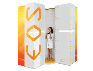
The EOS system is primarily used to assess patients with spine, hip and leg disorders. Because of the low radiation dose, EOS is ideal for progressive conditions that require frequent medical imaging, such as scoliosis. EOS is not typically used for injuries that can be evaluated with general radiography, such as broken bones in the arms, legs, hands or feet. Traditional X-rays are still the standard of care for diagnosing these injuries. For certain conditions, a 3D model of your child’s spine or lower limbs can be developed. 3D models provide our physicians with accurate measurements that can be used to make an informed diagnosis and develop treatment plans.
During the EOS exam, your child will sit or stand during the scan. The EOS exam will capture two images at the same time – one from the front and one from side. The scan takes approximately 20 seconds or less.
With EOS, our providers can make an informed diagnosis and develop treatment plans for your child’s spine, hip, or knee condition. Children’s is the only hospital in Nebraska offering EOS imaging.
The EOS imaging system is located in our Radiology Suite on the SPC 4th floor within the Orthopedic department.
-
Interventional Radiology (IR)
Pediatric interventional radiology (IR) is a medical field that specializes in minimally invasive diagnostic or therapeutic procedures using imaging guidance to diagnose and treat diseases.IR doctors guide small needles, catheters, and other small medical equipment into your child’s body through tiny incisions in the skin. Interventional radiologists use X-ray, CT scan, ultrasound, or MRI to see inside your child’s body to guide the instruments. Pediatric X-ray guided interventional procedures may require sedation or general anesthesia, depending on your child’s age and the type of procedure.
Our pediatric interventional radiologists are experts in these treatments and their follow-up in children, whose small size and special needs may require extra care. We are trained in special techniques appropriate for pediatric patients and use tools and imaging equipment created or modified for children.
Because some pediatric interventional procedures rely on x-ray technology, we have adapted our equipment and protocols to keep radiation exposure as low as reasonably achievable (the ALARA standard) during your child’s procedure.
Learn more about IR and the services we provide at Children’s in our IR Clinic.
-
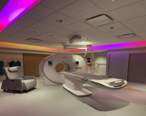
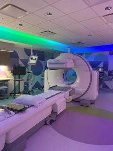
Magnetic Resonance Imaging (MRI)
Magnetic resonance imaging (MRI) is a procedure that uses a large magnet, radio-frequencies, and a computer to produce detailed images of body structures, including organs, tissues, and bones. It is used to help diagnose or monitor many types of medical conditions, such as heart defects or cancer. There is no radiation exposure during an MRI.
During the MRI, your child will lie on a narrow table that slides into the scanner, and will need to remain still throughout the whole scan. As long as we don’t need to use a sedative or anesthesia, you can stay in the room with your child during the scan.
For certain MRI scans, your child may need to receive contrast — a substance that’s taken either by mouth or injected directly into the body via an IV, and allows us to see the organ or tissue being studied more clearly. There are times your child will receive the contrast both orally and through an IV.
Staying still in an enclosed MRI machine can be difficult for anyone, including a child. It can also be a bit scary to be in a narrow machine that makes loud noises. We want our patients to be as comfortable as possible and remain still, so we can get the clearest images.
That’s why we have taken steps to make sure that our MRI scanners aren’t just child-friendly — they’re also fun.
Both of our scanners are decorated in a calming underwater theme on the outside and are outfitted with a unique TV/video system on the inside — something you won’t find in any other MRI scanner in Nebraska. Your child will wear headphones to hear the soundtrack of the movie, protect their ears from loud noises that the machine makes, and hear instructions from the MRI staff.
With a movie rolling and the loud noises of the machine being drowned out, many children can remain still and quiet during the 30- to 60-minute study without needing a sedative.
-
Nuclear Medicine/SPECT Studies
Nuclear medicine is a branch of radiology that allows us to not only see structures within the body, but also see how well they are functioning. It is used to diagnose diseases, injuries, or irregularities in the body, and assist in planning treatment.
For a nuclear medicine study, your child will be given a very small amount of radioactive material in the form of radiotracers. The radiotracers travel to the part of the body being studied. Usually, radiotracers are injected through an IV, but they can also be swallowed, inhaled, or delivered through a catheter (tube) placed into the bladder or stomach.
As radiotracers travel through the body, they emit invisible energy called gamma rays, which are then picked up by cameras. The cameras produce images of the movement and location of the radiotracer, which helps us assess how the structures in the body are functioning.
SPECT/CT or single photon emission computed tomography (SPECT)/computed tomography (CT) is a type of nuclear medicine scan that shows functional information and anatomical structures together. A SPECT/CT exam is usually ordered to diagnose or find out more about a specific disease state in the body. The SPECT portion of the exam reveals how organs, tissues or bones are functioning at the cellular level, and the CT portion of the exam provides information such as size, shape, and location. Combining these together enables physicians to accurately diagnose and identify various diseases and conditions within the body.
Nuclear medicine tests can last anywhere from 10 minutes to 4 hours, depending on the type of study. As long as your child does not need anesthesia, you will be allowed to stay in the room.
Your child will be exposed to a very small amount of radiation. Every dose is calculated specifically for your child, to ensure the smallest amount radiotracer is used to gain the best quality images. Since your child’s safety is our priority, we follow recommendations from the Image Gently® Campaign — a program from the Alliance for Radiation Safety in Pediatric Imaging that promotes X-ray safety measures specifically geared towards protecting children from radiation.
-
Ultrasound
An ultrasound uses sound waves to create pictures of organs. A handheld probe, called a transducer, sends the waves out at a frequency too high to be heard by human ears. These waves bounce off the organs like an echo, return to the transducer, and are converted into an electronic picture of the organs.
Your child’s physician might recommend an ultrasound for a number of reasons. The procedure can assess the size and location of abdominal organs and structures. It can also detect problems such as tumors (lumps of tissue), blood clots, infections, or excess fluid buildup in the abdomen.
During an ultrasound, we apply a clear, water-based gel to your child’s skin to allow for smooth movement, as well as to eliminate air between the skin and the transducer. The test is very safe — your child will not need to receive a contrast or be exposed to radiation.
Ultrasounds are painless, but your child may be a little uncomfortable about laying down for the entire length of the procedure, or feel nervous if they don’t understand what’s happening. Our technologist will explain everything to your child, from why we use the gel, to how they need to lay down, to what they’re seeing on the screen as the sound waves turn into pictures.
-
X-rays
X-rays are the most basic form of medical imaging. They are invisible beams of radiation that pass through the body, creating images of the bones and soft tissue. Children can have X-rays taken to look for broken bones, diagnose certain diseases like pneumonia, or spot signs of other serious medical conditions.
At Children’s, our X-rays are acquired in a digital format called digital radiography. This replaces traditional film, enhancing the image quality, lowering dose, and making X-ray images easily accessible as digital files.
Even though X-rays use radiation, they are safe. The amount of radiation is very small — about as much as your child would be exposed to naturally in their day-to-day life over the course of 10 days. Depending on the area being imaged, your child may also wear protective gear, such as an apron or lead “blanket”, which is specially designed to protect the body from radiation.
Safety is one of our top priorities, which is why we follow recommendations from the Image Gently® Campaign — a program from the Alliance for Radiation Safety in Pediatric Imaging that promotes X-ray safety measures specifically geared towards protecting children from radiation.
“Our experience was so amazing. The technologists were compassionate and friendly with my child. They helped him to understand what he had to do and explained each step. They made my son really enjoy his time in radiology.”
—Parent of Children’s Nebraska patient

Patient Education: Radiology Teaching Sheets
Learn about the most common radiology exams, and what you and your child can expect.
Our Pediatric Radiologists


What To Do Next
For Patients
Your child will need an order from a provider to schedule a radiology procedure. Once the order is placed, call 402-955-6799 to schedule the procedure. Take advantage of our offsite clinics to save yourself a trip to the hospital. Please call ahead, as not all studies can be completed offsite.
Information on imaging services offered at the Children’s Specialty Pediatric Clinic in Lincoln can be found here.
For Referring Providers
The Physicians’ Priority Line is your 24-hour link to pediatric specialists at Children’s for emergency and urgent consults, physician-to-physician consults, admissions, and transport services. Call 855-850-KIDS (5437).
Learn more about referring patients.